What Are The Indications Of Inferior Calcaneal Spur

Overview
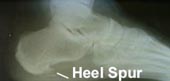
The plantar fascia is connective tissue on the sole of your foot. When the arch of the foot is not properly supported, the plantar fascia can stretch and pull away from the heel area. When the plantar fascia pulls away from the heel, calcium deposits form in its absence. These calcium deposits are called heel spurs and can be very painful.
Causes
Heel spurs develop in some people that have a condition called plantar fasciitis, inflammation of the plantar fascia. Heel spurs form when the plantar fascia separates from the calcaneus. An abnormal bone growth, a hook-like spur, forms from calcium deposits that grow at the site of inflammation. Heel spurs are more common in middle-aged adults and people that have had plantar fasciitis for a long time. People with flat feet or high arches are vulnerable to heel spurs. Women who wear high-heeled shoes are more susceptible, as well.
Symptoms
More often than not, heel spurs have no signs or symptoms, and you don?t feel any pain. This is because heel spurs aren?t pointy or sharp pieces of bone, contrary to common belief. Heel spurs don?t cut tissue every time movement occurs; they?re actually deposits of calcium on bone set in place by the body?s normal bone-forming mechanisms. This means they?re smooth and flat, just like all other bones. Because there?s already tissue present at the site of a heel spur, sometimes that area and the surrounding tissue get inflamed, leading to a number of symptoms, such as chronic heel pain that occurs when jogging or walking.
Diagnosis
Your doctor will discuss your medical history and will examine your foot and heel for any deformities and inflammation (swelling, redness, heat, pain). He/she will analyze your flexibility, stability, and gait (the way you walk). Occasionally an x-ray or blood tests (to rule out diseases or infections) may be requested.
Non Surgical Treatment
Some heel spurs do require surgery, however surgery is a last resort. In most cases the patients underlying foot problem needs to be addressed, such as Over Pronation and Over Supination and Heel Pain Treatment Options need to be implemented if Plantar Fasciitis and Achilles Tendonitis are still an ongoing concern. Your best treatment is always prevention.
Surgical Treatment
Heel spur surgery should only be considered after less invasive treatment methods have been explored and ruled insufficient. The traditional surgical approach to treating heel spurs requires a scalpel cut to the bottom of the food which allows the surgeon to access the bone spur. Endoscopic plantar fasciotomies (EPF) involve one or two small incisions in the foot which allow the surgeon to access and operate on the bone spur endoscopically. Taking a surgical approach to heel spur treatment is a topic to explore with a foot and ankle specialist.
Prevention
You can help prevent heel spur symptoms from returning by wearing the proper shoes. Customized orthotics and insoles can help relieve pressure. It is important to perform your exercises to help keep your foot stretched and relaxed.
What Causes Bursitis Of The Feet?
Overview
Achilles bursitis is one of those injuries that can really bring down the quality of your life. Anyone, young or old, can suffer from this injury, and if you're active this condition will keep you from doing the things you love to do. It will even start interrupting any of your normal daily tasks and make living life harder than it really needs to be. Fortunately for you, professional athletes have had access to state of the art treatment therapies for years that allow them to heal more quickly and completely than you or I. This is why athletes that have a serious heel bursitis injury can often get back in the game in a matter of weeks while you could suffer for months or even years (in chronic cases).
Causes
If the posterior-superior portion of the heel has an abnormally large bony prominence protruding from it (called a Haglund's Deformity), in some instances it may rub against the Achilles Tendon. When this occurs, the bursa between the bone and the tendon will become inflamed, swollen, and painful. This condition is called Retrocalcaneal Bursitis. The presence of a Haglund's Deformity does not insure that these problems will occur. In order for these problems to occur, the heel and foot must be tilted in such a way as to actually force this bony prominence into the bursa and tendon.
Symptoms
Your feet are extremely resilient and are designed to stand up to the pressures of day-to-day living. In some cases, though, foot structures may break down when subjected to chronic stress associated with prolonged periods of weight-bearing activity on concrete, asphalt, or other hard surfaces (especially when your footwear does not allow for appropriate weight distribution). Foot problems, including infracalcaneal bursitis, are often exacerbated by poorly designed footwear, and pressure, impact, and shear forces can damage your feet over time. Bursal sacs are intended to minimize this damage, but sometimes the bursa itself becomes inflamed.
Diagnosis
Like all other forms of bursitis, initially the physician will take down the history of symptoms experienced by the patient, this will be followed by a detailed physical examination which involves checking for inflammation signs like pain, redness, and warmth of the heel area. The physician might examine further by moving the ankle a little to determine the exact location of pain. Further diagnostic tests including x-ray, bone scans, and MRI scan might be suggested if required.
Non Surgical Treatment
You should rest from all activities that cause pain or limping. Use crutches/cane until you can walk without pain or limping. Ice. Place a plastic bag with ice on the foot for 15-20 minutes, 3-5 times a day for the first 24-72 hours. Leave the ice off at least 1 1/2 hours between applications. Compression. Lightly wrap an elastic bandage from the toes to mid calf, using even pressure. Wear this until swelling decreases. Loosen the wrap if your toes start to turn blue or feel cold. Elevate. Make sure to elevate the ankle above heart level. To improve symptoms of plantar calcaneal bursitis after the acute phasetry the baked bean tin stretch, using a baked bean tin roll the foot backwards and forwards. 2 minutes in the morning before putting the foot to the floor. 5-10 minutes every evening. Contrast foot baths. 10 minutes warm water. 10 minutes cool water morning and evening (morning may be missed if time is restricted). Stretches. Start with 10 stretches per day, holding the stretch for 30 seconds, then relax and then repeat. Continue this stretch daily until you can no longer feel it pulling on the heel, then progress to stretch. Do 10 per day holding for 30 seconds per stretch. When you can no longer feel it pulling on the heel proceed to stretches. Do 10 per day holding for 30 seconds on every stretch.
Surgical Treatment
Surgery is rarely need to treat most of these conditions. A patient with a soft tissue rheumatic syndrome may need surgery, however, if problems persist and other treatment methods do not help symptoms.
Hammertoe Pain
 Overview
A hammertoes is a deformity of the second, third or fourth toes in which the main toe joint is bent upward like a claw. Initially, hammertoes are flexible and can be corrected with simple measures. Left untreated, they can become fixed and require surgery. Hammertoe results from shoes that don?t fit properly or a muscle imbalance, usually in combination with one or more other factors. Muscles work in pairs to straighten and bend the toes. If the toe is bent and held in one position long enough, the muscles tighten and can?t stretch out.
Causes
Hammer toe is commonly caused by wearing shoes that are too narrow, tight or short on a regular basis. By doing so, your toe joints are forced into odd position. Over time, the tendons and muscles in your toe become shorter and cause it to bend. You can suffer a hammer toe if you have diabetes and the disease is worsening. If this occurs, you should contact your doctor right away. Arthritis can also cause hammer toes. Because your toe muscles get out of balance when you suffer from this joint disorder, tendons and joints of your toes are going to experience a lot of pressure.
 Symptoms
Symptoms include sharp pain in the middle of the toe and difficulty straightening the toe. People with hammertoe may also develop blisters, which are fluid-filled pockets of skin, because the bent toe is likely to rub against the inside of a shoe. This increased friction may also lead to calluses, which are areas of thickened skin, and corns, which are hard lumps that may form on or between toes. Symptoms may be minor at first, but they can worsen over time.
Diagnosis
Hammer toes may be easily detected through observation. The malformation of the person's toes begin as mild distortions, yet may worsen over time - especially if the factors causing the hammer toes are not eased or removed. If the condition is paid attention to early enough, the person's toes may not be permanently damaged and may be treated without having to receive surgical intervention. If the person's toes remain untreated for too long, however the muscles within the toes might stiffen even more and will require invasive procedures to correct the deformity.
Non Surgical Treatment
Hammer toes usually get progressively worse over time, especially if you avoid seeking care. Not all cases are the same, so it is important to get your podiatrist or foot surgeon to evaluate your condition so that you can get the treatment you need as soon as possible. Your treatment options will vary depending on the severity of your hammer toe. You may not require surgery to treat your hammer toe. Your doctor may suggest one of these less invasive measures. Instead of wearing shoes that are too high or too short, wear comfortable shoes that have plenty of room and are flat or low-heeled. Your doctor can prescribe pads that will prevent your corns or calluses from getting irritated. Avoid over-the-counter medicated pads, as they contain Hammer toes acid that can worsen your condition. An orthotic device can be customized to fit your shoe and foot. It can help control your tendon and muscle imbalance, which in turn may ease your pain. NSAIDS (nonsteroidal anti-inflammatory drugs) such as ibuprofen can reduce inflammation. By relieving swelling in your toe joint, you can alleviate your pain. Splints or small straps can be placed on your toe by a foot surgeon to realign your bent toe. Applying ice packs wrapped in cloth on your hammer toe can reduce inflammation and swelling. Gently massaging your toes can assist in alleviating your pain caused by hammer toes. Try exercises that stretch your feet as these can help restore your muscle balance. A simple exercise that can help is to pick up a cloth or small object from the floor by curling your toes. This action will help your feet and toes by stretching them.
Surgical Treatment
If your hammer, claw, or mallet toe gets worse, or if nonsurgical treatment does not help your pain, you may think about surgery. The type of surgery you choose depends on how severe your condition is and whether the toe joint is fixed (has no movement) or flexible (has some movement). A fixed toe joint often requires surgery to be straightened. A flexible toe joint can sometimes be straightened without surgery. Surgery choices include Phalangeal head resection (arthroplasty), in which the surgeon removes part of the toe bone. Joint fusion (arthrodesis), in which the surgeon removes part of the joint, letting the toe bones grow together (fuse). Cutting supporting tissue or moving tendons in the toe joint. How well surgery works depends on what type of surgery you have, how experienced your surgeon is, and how badly your toes are affected.
Everything You Need To Know Regarding Bunions
Overview
 When the big toe is misaligned, it creates an unnatural bump on the inside of the forefoot (see image below). In addition to being unsightly, the lump or bunion can cause pain and make buying shoes difficult. Some people are born predisposed to bunions. But most people acquire them over time from ill-fitting footwear that squeezes the big toe inward toward the other toes. About four out of five bunion pain patients are female, a near perfect fit for the bad shoe theory. The good news, Bunions can be corrected, often with better-fitting shoes and custom orthotics. There are also some cases that cause severe bunion pain and do not respond to conservative treatment. For those patients, bunion surgery called bunionectomy may be necessary.
Causes
It is thought that the primary cause of bunions is a mechanical instability in the big toe joint. There are a number of different reasons as to why this may happen. Bunions tend to run in families so a person with a family history of bunions has an increased risk of developing them. Factors that are known to increase the risk of bunions include wearing improperly fitting shoes (particularly narrow and/or high-heeled shoes). Certain arthritic conditions and ligament disorders. Age (the incidence of bunions increases with age). Being flatfooted with feet that roll inwards (over pronation). Past injury (trauma) to the foot. Nerve conditions affecting the foot. Bunions most commonly affect women (approximately 90% of cases) and are less common in people who do not regularly wear shoes.
Symptoms
The signs and symptoms of a bunion include a bulging bump on the outside of the base of your big toe, swelling, redness or soreness around your big toe joint, Thickening of the skin at the base of your big toe, Corns or calluses, these often develop where the first and second toes overlap, persistent or intermittent pain, restricted movement of your big toe. Although bunions often require no medical treatment, see your doctor or a doctor who specializes in treating foot disorders (podiatrist or orthopedic foot specialist) if you have persistent big toe or foot pain, a visible bump on your big toe joint, decreased movement of your big toe or foot, difficulty finding shoes that fit properly because of a bunion.
Diagnosis
Your doctor will ask questions about your past health and carefully examine your toe and joint. Some of the questions might be: When did the bunions start? What activities or shoes make your bunions worse? Do any other joints hurt? The doctor will examine your toe and joint and check their range of motion. This is done while you are sitting and while you are standing so that the doctor can see the toe and joint at rest and while bearing weight. X-rays are often used to check for bone problems or to rule out other causes of pain and swelling. Other tests, such as blood tests or arthrocentesis (removal of fluid from a joint for testing), are sometimes done to check for other problems that can cause joint pain and swelling. These problems might include gout , rheumatoid arthritis , or joint infection.
Non Surgical Treatment
Bunion pain can be successfully managed in the vast majority of cases by switching to shoes that fit properly and don't compress the toes. Your orthopaedic surgeon can give you more information about proper shoe fit and the types of shoes that would be best for you. Follow these general points of shoe fit. Do not select shoes by the size marked inside the shoe. Sizes vary among shoe brands and styles. Judge the shoe by how it fits on your foot. Select a shoe that conforms as nearly as possible to the shape of your foot. Have your feet measured regularly. The size of your feet change as you grow older. Have both feet measured. Most people have one foot larger than the other. Fit to the largest foot. Fit at the end of the day when your feet are the largest. Stand during the fitting process and check that there is adequate space (3/8" to 1/2") for your longest toe at the end of each shoe. Make sure the ball of your foot fits well into the widest part (ball pocket) of the shoe. Do not purchase shoes that feel too tight, expecting them to "stretch" to fit. Your heel should fit comfortably in the shoe with a minimum amount of slippage. Walk in the shoe to make sure it fits and feels right. (Fashionable shoes can be comfortable.) Some shoes can be modified by stretching the areas that put pressure on your toes. Splints to reposition the big toe and orthotics (special shoe inserts shaped to your feet) also may relieve pain. For bunions caused by arthritis, medications can be prescribed to reduce pain and swelling. 
Surgical Treatment
For those whose bunions cause persisting pain, a surgical operation is considered for correction of the bunion. The surgical operation to correct a bunion is referred to as a bunionectomy. Surgical procedures can correct deformity and relieve pain, leading to improved foot function. These procedures typically involve removing bony growth of the bunion while realigning the big toe joint. Surgery is often, but not always, successful; failure to relieve pain can result from the big toe moving back to its previous deviated position even after surgery. However, proper footwear and orthotics can reduce the chances of surgical failure.
Prevention
If these exercises cause pain, don't overdo them. Go as far as you can without causing pain that persists. This first exercise should not cause pain, but is great for stimulating blood and lymphatic circulation. Do it as often as you can every day. Only do this exercise after confirming it is OK with your doctor. Lie on your back and lift up your legs above you. Wiggle your toes and feet. Eventually you may be able to rapidly shake your feet for a minute at a time. Use your fingers to pull your big toe into proper alignment. Stretch your big toe and the rest of your toes. Curl them under for 10 seconds, then relax and let them point straight ahead for 10 seconds. Repeat several times. Do this at least once a day, and preferably several times. Flex your toes by pressing them against the floor or a wall until they are bent back. Hold them for 10 seconds, then release. Repeat several times. Grip with your toes. Practice picking up an article of clothing with your toes, dropping it, and then picking it up again. Warm water. Soak your feet for 20 minutes in a bowl of warm water. Try doing the foot exercises while soaking, and also relax and rest your feet. Epsom salts. Add it to your warm foot bath soak.
Understand Overpronation Of The Foot
Overview
Over-pronation describes a characteristic gait wherein a lack of sufficient support from the arch of the foot causes it to roll inwards as weight is placed on it. The long arch of the foot is actually a very important structure in terms of our gait and how we walk, it is responsible for ensuring that as our weight rolls from the heel to the balls of our feet it does so in a straight line that doesn?t place undue stress on the ankle or knees. Unfortunately when a person?s arch is not pronounced enough, or even simply not there (a condition called flat footedness), there isn?t enough arch support to maintain a healthy, forward motion of the foot.
Causes
Overpronation often occurs in people with flat feet, whose plantar fascia ligament is too flexible or too long, and therefore unable to properly support the longitudinal arch of the foot. People tend to inherit the foot structure that leads to overpronation. In a normal foot the bones are arranged so that two arches are formed, the longitudinal and the transverse. Ligaments hold all the bones in their correct positions, and tendons attach muscles to bones. If the bones are held together too loosely, they will tend to move inwards as this is the easiest direction for them to go. Over time the soft tissue structures will adjust to the misalignment and the foot will become permanently over-flexible, with a flat arch.
Symptoms
It is important to note that pronation is not wrong or bad for you. In fact, our feet need to pronate and supinate to achieve proper gait. Pronation (rolling inwards) absorbs shock and supination (rolling outwards) propels our feet forward. It is our body?s natural shock-absorbing mechanism. The problem is over-pronation i.e. the pronation movement goes too deep and lasts for too long, which hinders the foot from recovering and supinating. With every step, excess pronation impedes your natural walking pattern, causing an imbalance in the body and consequent excessive wear and tear in joints, muscles and ligaments. Some common complaints associated with over-pronation include Heel Pain (Plantar Fasciitis) ,Ball of foot pain, Achilles Tendonitis, Shin splints, Knee Pain, Lower Back Pain.
Diagnosis
When sitting, an over-pronating foot appears quite normal, i.e. showing a normal arch with room under the underside of the foot. The moment you get up and put weight on your feet the situation changes: the arches lower and the ankle slightly turns inwards. When you walk or run more weight is placed on the feet compared to standing and over-pronation will become more evident. When walking barefoot on tiles or timber floors over-pronation is more visible, compared to walking on carpet or grass.
Non Surgical Treatment
If a young child is diagnosed with overpronation braces and custom orthotics can be, conjunction with strengthening and stretching exercises, to realign the bones of the foot. These treatments may have to continue until the child has stopped growing, and orthotics may need to be worn for life in order to prevent the foot reverting to an overpronated state. Wearing shoes that properly support the foot, particularly the arch, is one of the most effective treatments for overpronation. Custom-made orthotic inserts can also be very beneficial. They too support the arch and distribute body weight correctly throughout the foot. Motion-control shoes that prohibit pronation can be worn, so may be useful for those with severe overpronation. One good treatment is to walk barefoot as often as possible. Not relying on shoes to support the arch will encourage proper muscle use. Practicing yoga can help to correct poor posture and teach you how to stand with your weight balanced evenly across the whole foot.
Prevention
Firstly, a thorough and correct warm up will help to prepare the muscles and tendons for any activity or sport. Without a proper warm up the muscles and tendons around your feet, ankles and lower legs will be tight and stiff. There will be limited blood flow to the lower legs, which will result in a lack of oxygen and nutrients for those muscles. Click here for a detailed explanation of how, why and when to perform your warm up. Secondly, flexible muscles are extremely important in the prevention of most ankle and lower leg injuries. When muscles and tendons are flexible and supple, they are able to move and perform without being over stretched. If however, your muscles and tendons are tight and stiff, it is quite easy for those muscles and tendons to be pushed beyond their natural range of motion. To keep your muscles and tendons flexible and supple, it is important to undertake a structured stretching routine.
Calcaneal Apophysitis Physiotherapy
Overview
Foot pain is not only limited to grown-ups. Often, healthy and active children will complain of pain in one or both heels shortly after running, engaging in sports or even walking. The pain is centered at the back of, or under the heel. The cause of heel pain in children is usually a condition called calcaneal apophysitis, normally reported by 8 to 14 year olds.
Causes
Sever condition is caused by sprain injury where the Achilles tendon attaches to the calcaneus bone at the back of the heel. Sever condition occurs in adolescent or older children, particularly active boys. It can be very painful. It is one of those conditions commonly referred to as "growing pains." Patients are evaluated for signs of conditions that can mimic Sever condition, such as ankylosing spondylitis and other forms of arthritis. Usually Sever condition is self-limited; that is, it disappears as the child ages.
Symptoms
This syndrome can occur unilaterally or bilaterally. The incidence of bilaterally is approximately 60%. Common signs and symptoms include posterior inferior heel pain (over the medial and lateral surface of the bone). Pain is usually absent when the child gets up in the morning. Increased pain with weight bearing, running or jumping (= activity-related pain). The area often feels stiff. The child may limp at the end of physical activity. Tenderness at the insertion of the tendons (= an avascular necrosis of the arthropathy). Limited ankle dorsiflexion range secondary to tightness of the Achilles tendon. Hard surfaces and poor-quality or worn-out athletic shoes contribute to increased symptoms. The pain gradually resolves with rest. Reliability or validity of methods used to obtain the ankle joint dorsiflexion or biomechanical malalignment data are not commented upon, thus reducing the quality of the data. Although pain and limping are mentioned as symptomatic traits, there have been no attempts to quantify the pain or its effect on the individual.
Diagnosis
All medical diagnosis should be made by taking a full history, examining the patient then performing investigations. The problem usually occurs in boys who are going through or have just gone through a growth spurt; one or both heels may be affected. Initially the pain may be intermittent occurring only during or after exercise. As the problem gets worse, pain may be present most of the time. There may be swelling over the back of the heel and this area is painful if touched or knocked. On examination the patient often has flat feet, very tight legs muscles especially the gastrocnemius.
Non Surgical Treatment
Treatment depends on the severity of the condition, but may include relative rest and modified activity, a physiotherapist can help work out what, and how much, activity to undertake. Cold packs, apply ice or cold packs to the back of the heels for around 15 minutes after any physical activity, including walking. Shoe inserts, small heel inserts worn inside the shoes can take some of the traction pressure off the Achilles tendons. This will only be required in the short term. Medication, pain-relieving medication may help in extreme cases, but should always be combined with other treatment and following consultation with your doctor). Anti-inflammatory creams are also an effective management tool. Splinting or casting, in severe cases, it may be necessary to immobilise the lower leg using a splint or cast, but this is rare. Time, generally the pain will ease in one to two weeks, although there may be flare-ups from time to time. Correction of any biomechanical issues, a physiotherapist can identify and discuss any biomechanical issues that may cause or worsen the condition. Education on how to self-manage the symptoms and flare-ups of Sever?s disease is an essential part of the treatment.
Prevention
The chances of a child developing heel pain can be reduced by avoiding obesity. Choosing well-constructed, supportive shoes that are appropriate for the child?s activity. Avoiding or limiting wearing of cleated athletic shoes. Avoiding activity beyond a child?s ability.
Tibial Posterior Tendon Dysfunction Stages
Overview
Adult-acquired flatfoot is a challenging condition to treat. It is defined as a symptomatic, progressive deformity of the foot caused by loss of supportive structures of the medial arch. It is becoming increasingly frequent with the aging population and the obesity epidemic. Patients commonly try to lose weight by exercising to improve the condition. This often leads to worsening of symptoms and progression of the disorder. Early recognition of this complex disorder is essential, if chronic pain and surgery are to be avoided. 
Causes
Several risk factors are associated with PTT dysfunction, including high blood pressure, obesity, diabetes, previous ankle surgery or trauma and exposure to steroids. A person who suspects that they are suffering from PTT dysfunction should seek medical attention earlier rather than later. It is much easier to treat early and avoid a collapsed arch than it is to repair one. When the pain first happens and there is no significant flatfoot deformity, initial treatments include rest, oral anti-inflammatory medications and, depending on the severity, a special boot or brace.
Symptoms
Pain and swelling around the inside aspect of the ankle initially. Later, the arch of the foot may fall (foot becomes flat), this change leads to walking to become difficult and painful, as well as standing for long periods. As the flat foot becomes established, pain may progress to the outer part of the ankle. Eventually, arthritis may develop.
Diagnosis
Observation by a skilled foot clinician and a hands-on evaluation of the foot and ankle is the most accurate diagnostic technique. Your Dallas foot doctor may have you do a walking examination (the most reliable way to check for the deformity). During walking, the affected foot appears more pronated and deformed. Your podiatrist may do muscle testing to look for strength deficiencies. During a single foot raise test, the foot doctor will ask you to rise up on the tip of your toes while keeping your unaffected foot off the ground. If your posterior tendon has been attenuated or ruptured, you will be unable to lift your heel off the floor. In less severe cases, it is possible to rise onto your toes, but your heel will not invert normally. X-rays are not always helpful as a diagnostic tool for Adult Flatfoot because both feet will generally demonstrate a deformity. MRI (magnetic resonance imaging) may show tendon injury and inflammation, but can?t always be relied on for a complete diagnosis. In most cases, a MRI is not necessary to diagnose a posterior tibial tendon injury. An ultrasound may also be used to confirm the deformity, but is usually not required for an initial diagnosis.
Non surgical Treatment
Because of the progressive nature of PTTD, early treatment is advised. If treated early enough, your symptoms may resolve without the need for surgery and progression of your condition can be arrested. In contrast, untreated PTTD could leave you with an extremely flat foot, painful arthritis in the foot and ankle, and increasing limitations on walking, running, or other activities. In many cases of PTTD, treatment can begin with non-surgical approaches that may include. Orthotic devices or bracing. To give your arch the support it needs, your foot and ankle surgeon may provide you with an ankle brace or a custom orthotic device that fits into the shoe. Immobilization. Sometimes a short-leg cast or boot is worn to immobilize the foot and allow the tendon to heal, or you may need to completely avoid all weight-bearing for a while. Physical therapy. Ultrasound therapy and exercises may help rehabilitate the tendon and muscle following immobilization. Medications. Nonsteroidal anti-inflammatory drugs (NSAIDs), such as ibuprofen, help reduce the pain and inflammation. Shoe modifications. Your foot and ankle surgeon may advise changes to make with your shoes and may provide special inserts designed to improve arch support. 
Surgical Treatment
For those patients with PTTD that have severe deformity or have not improved with conservative treatments, surgery may be necessary to return them to daily activity. Surgery for PTTD may include repair of the diseased tendon and possible tendon transfer to a nearby healthy tendon, surgery on the surrounding bones or joints to prevent biomechanical abnormalities that may be a contributing factor or both.